Slashdot Mirror
Domain: prefrontal.org
Stories and comments across the archive that link to prefrontal.org.
Comments · 11
-
This applies mostly to medicine and social science
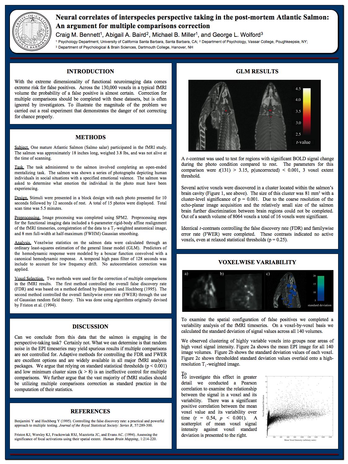
This applies to mostly medicine and social science see John Ioannidis's research paper "Why Most Published Research Findings Are False" : http://journals.plos.org/plosm... It seems to me the sciences that deal with statistical p-value significance are all subject to false published research findings , for instance, see Craig Bennet's "Neural correlatates of interspecies perspectitve taking in post-mortem Salmon : An argument for multiple comparisons corrections". http://prefrontal.org/files/po... The paper is a deadpan gag and a veiled attack on sloppy methodology among neuroimaging researchers. Also, researchers run the Baltimore Stockbroker scam : https://somemathematicalmusing... When they selectively choose not to publish certain results in favor of other ones etc... So on and so forth etc...
-
Salmon inspired neural networks
They could train their deep learning networks based on research done on brain of dead salmon reacting to human emotions...
-
Re:Or other things
more research is needed to determine whether similar findings could be replicated in people of other faiths, such as Catholics or Muslims.
Or favorite sports teams, or social movements, or fandoms, or whatever else makes people tick.
Or dead salmon.
-
Oh dear...
what a primitive definition of 'free will'. One day we might come up with a realistic model of what it is and be able to create experiments that really test it. In the meantime it might be worth taking a look at http://prefrontal.org/files/posters/Bennett-Salmon-2009.pdf - there is a growing body of evidence that fMRI correlates to brain activity are often questionable.
-
Re:Why can't they give us the MRI image instead?
We have PET scan, we have MRI, we have the technology to do virtual 3D slicing.
Exactly! I just can't wait for the article titled "Neural correlates of perspective taking in the post-mortem Einstein's brain"
(this is to say: what the hell is one expected to find in a brain dead for more than half a century?!)
-
I'm sure a post mortem salmon will do
http://prefrontal.org/files/posters/Bennett-Salmon-2009.pdf
When it comes to fMRI studies, I always remember the story of a dead salmon in an fMRI scanner, that was shown a series of photographs depicting human individuals in social situations. The salmon was asked to determine what emotion the individual in the photo must have been experiencing.
Of course, it was a resounding success! And now SCIENCE knows where in the brain of a dead salmon, the mental process to evaluate human emotions occurs.
-
I'm sure a post mortem salmon will do
http://prefrontal.org/files/posters/Bennett-Salmon-2009.pdf
When it comes to fMRI studies, I always remember the story of a dead salmon in an fMRI scanner, that was shown a series of photographs depicting human individuals in social situations. The salmon was asked to determine what emotion the individual in the photo must have been experiencing.
Of course, it was a resounding success! And now SCIENCE knows where in the brain of a dead salmon, the mental process to evaluate human emotions occurs.
-
Salmon
So, did they also try a dead salmon?
Just think what this could mean: well-educated zombies!
-
Re:I've Seen It All
I'm sure you've seen this, yes?
-
Any questions?
Hey guys - I am the first author of the Salmon poster. If you have any questions that you would like us to answer then post it as a reply below and I will do my best to respond as soon as I can.
You can find some more information on the poster at the following link:
http://prefrontal.org/blog/2009/06/atlantic-salmon-index/Best ~ Craig Bennett
-
Re:"These images are not snapshots"? No kidding.
...and yet, it does. It's become so routine, so reliable, so well-understood and well-controlled, that doctors and researchers know they can rely on it as a matter of course. They still have to be aware of the errors and distortions that can arise, but that's true of every imaging or monitoring system, all the way down to the stethoscope and the fever thermometer.
The problem with the activation maps is precisely that one is NOT looking at an image, so there's no way to fine tune the algorithms. Therefore, fMRI is NOT well understood in the way that CT or MRI are.
Consider that in imaging, you have the luxury of comparing the output of a brain scan to the known physical structure of the brain. Is there a hippocampus? No? Well then it didn't work, go back and fiddle until you can show me a hippocampus.
In fMRI, apart from low level sensory corticies (where visual field mapping techniques can reproduce broad level retinotopic maps), researchers are operating in a vacuum in which there is no hard and fast error signal to fine tune the methods.
Science has to proceed very cautiously in such a situation. This is particularly true when one has hundreds of thousands of voxels to sift through because it's easy to find any pattern in noise, if you have enough noise.
So I would argue that fMRI offers a very different set of challenges compared to MRI and CT scans, and therefore it's very important to keep a sharp, critical eye on the statistics used, as these authors are doing.
To illustrate this point further, here is a link to a poster in which someone put a dead salmon into a magnet and found that (in the absence of proper statistical controls) its decomposing brain was apparently reacting to the emotional content of pictures: